Asyn Fibrils
Aggregation of misfolded proteins is a feature common to many neurodegenerative diseases. Parkinson’s disease is a progressive movement disorder which is characterized neuropathologically by the presence of intraneuronal Lewy Bodies (LB) and Lewy Neurites (LN). Misfolded and aggregated alpha-synuclein (fibrillar alpha-synuclein) is the major component of LB’s and LN’s.
Synthetic alpha-synuclein fibrils (murine and human PFF) are capable of ‘seeding’ and propagating alpha synuclein pathology in both alpha-synuclein transgenic and non-transgenic (WT) neuronal cultures and mice (Luk, et al., 2012a; Luk, et al., 2012b; Volpicelli-Daley, et al., 2014).
To characterize the extensive early alpha-synuclein pathology and the reduction in striatal dopamine levels in 30 days post synthetic alpha-synuclein fibril injected WT mice, PsychoGenics conducted a range of assessments from standard and proprietary behavioral paradigms, microdialysis, electrophysiology and immune-histochemistry to further characterize the model.
Coordination Deficit Seen at 30 and 90 Days Post Injection of Alpha-Synuclein Pre-Formed Fibrils [PFF]
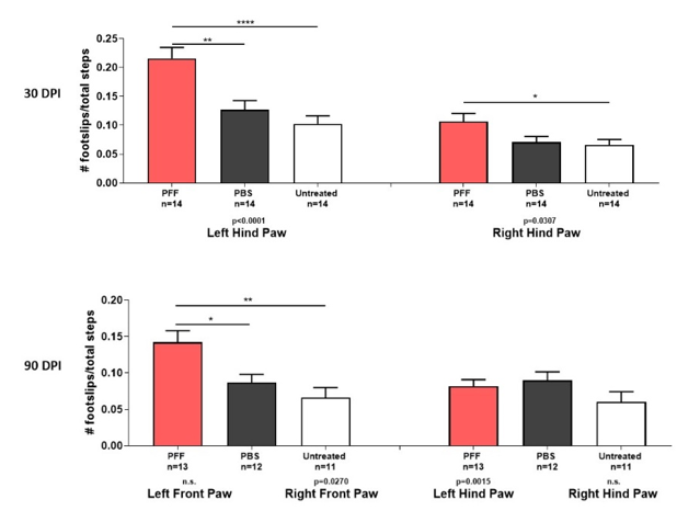
Figure 1: Latency to turn and number of footslips on tapered balance beam at 30 and 90 days post unilateral striatal inoculation of PFF. Mice were required to successfully complete a minimum of two beam traversals in a testing session (max of 2 minutes each session) in order to be scored for number of footslips. The most significant deficits in footslips are observed in the contralateral hind limb – the Left Hind Paw. [Sample size=14, Male B6C3F1/J mice (8-10)].
Reduction in Average Wire Hang-Time in Mice Injected with PFF’s at 30 and 90 Days Post-Injection
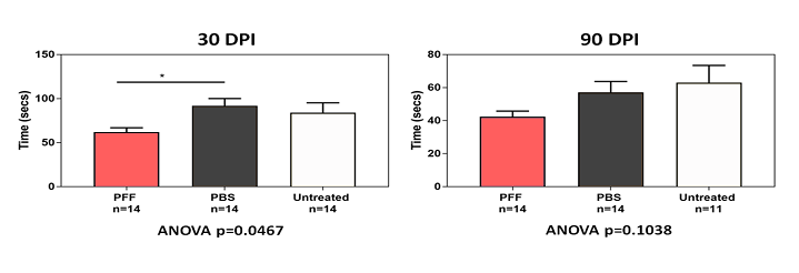
Figure 2: Shorter latency to hang on the wire hang test at 30 and 90 days post unilateral striatal inoculation of alpha-synuclein preformed fibrils. Mice were allowed to grip the steel grid for as long as possible. Mice were given three trials with an ITI of 2-3minutes. [Sample size=14, Male B6C3F1/J mice (8-10 weeks) mice per treatment *** P<0.05, Unpaired t-test].
NeuroCube® Analysis Reveals Progressive Gait Impairments in Stride and Swing Duration and in Forelimb Paw Placement 90 Days Post-Injection of PFFs
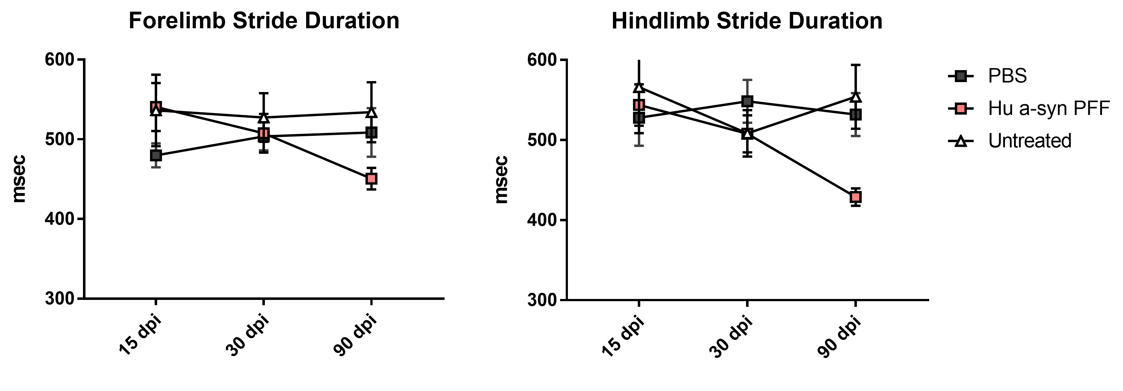
Figure 3: Gait assessment using PsychoGenics proprietary gait platform: NeuroCube® employing computer vision to detect changes in gait geometry and gait dynamics. Mice were tested for 5 minutes in a rectangular Neurocube® chamber where mice were allowed move freely back and forth through the rectangular walkway. Complex bioinformatics algorithms are employed to detect subtle gait phenotypes. [Sample size=14 Male B6C3F1/J mice (8-10 weeks) mice per treatment *** P<0.05, Unpaired t-test].
Intrastriatal Injection of Human PFFs Reduced Glutamatergic Transmission in Dorsal Striatum Spiny Projection Neurons (SPNs)
Miniature excitatory postsynaptic currents (mEPSC) frequency measures presynaptic release or changes in the number of synapses, whereas changes in the amplitude translates to alterations in glutamate receptor function in the postsynaptic compartment.
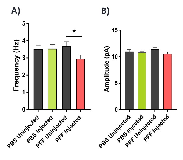
Figure 4: Analysis of action potential-independent glutamatergic transmission of striatal SPNs in the dorsal striatum using whole-cell patch clamp electrophysiology in brain slices. 2-3 month-old wild-type mice received an unilateral intrastriatal injection of either phosphate buffered saline (PBS) or human α-synuclein PFFs and aged for 180 days. Brain slices were then prepared from PBS versus PFF injected mice and (mEPSCs) were recorded in the presence of 1αM tetrodotoxin and 40 αM picrotoxin (n=30 neurons from 6 mice per group). (A) Frequency of mEPSCs was significantly reduced in the hemisphere injected with PFF mice, whereas in panel (B) the amplitude of mEPSCs was unaltered. The robust alterations in mEPSC frequency suggests a potential loss of glutamatergic synapses or decreased probability of transmitter release in striatal SPNs exposed to PFFs.