Preclinical Models of Traumatic Brain Injury
Traumatic brain injury (TBI) has been called a silent epidemic. The United States Centers for Disease Control and Prevention reports that over 2.5 million TBIs occurred in the United States in 2010 alone. Pharmacotherapies targeted at mitigating early post-traumatic secondary degenerative pathology such as inflammation, oxidative stress and excitotoxicity, as well as therapies to promote brain plasticity in order to restore function continue to require preclinical testing. PsychoGenics conducts a broad spectrum of post-traumatic behavioral tests to comprehensively assess the preclinical efficacy of novel therapies for TBI.
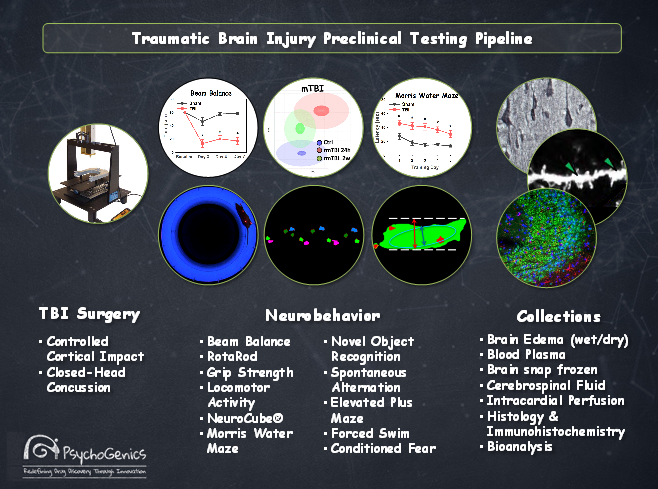
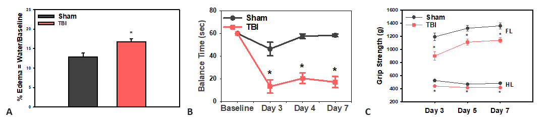
Figure 1: Early pathology following TBI. (A) Increased brain edema at 48 hours post-trauma measured by wet-dry weight method. Post-traumatic motor deficits in beam balance (B) and grip strength (C).
SmartCube® and NeuroCube® Systems
The SmartCube® System (SC) is a high content platform that measures behavioral responses and the NeuroCube® System (NC) is an automated gait analysis platform that measures gait geometry, gait dynamics, body movements, paw pressure and paw position among other gait features.
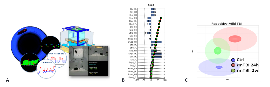
Figure 2: (A) NeuroCube® testing and image processing of locomotor metrics and SmartCube® schematic showing the three camera views with classification. (B) Combined gait features ranked by magnitude of discrimination between Sham and TBI. (C) SmartCube® phenotype of repetitive mild TBI at 24 hrs. and partial recovery at 2 weeks after rmTBI.
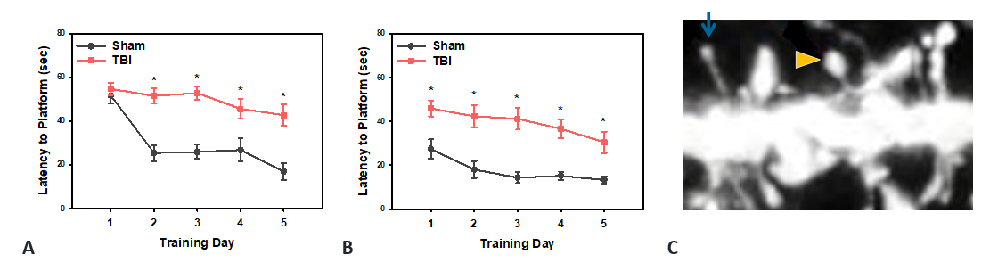
Figure 3: Persistent cognitive deficits in Morris Water Maze. (A) Escape latency from water maze at 3 weeks post-trauma. (B) Morris Water Maze reversal learning at 3 months post-trauma. (C) Morphometric analysis of dendritic spines (arrow marks thin spine, arrowhead marks mushroom spine). TBI causes loss of dendritic spine density.
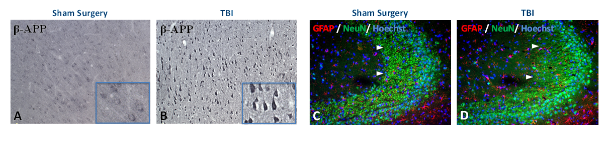
Figure 4: Beta amyloid precursor protein visibly accumulates in neuronal perikarya at 3 weeks post-trauma (B) whereas sham surgical controls (A) exhibit only faint immunostaining for β-APP. Immunofluorescence staining for glial fibrillary acidic protein (red) and NeuN (green) reveals neuronal loss in CA3 region of hippocampus following neurotrauma (arrowheads in C vs. D).